


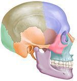
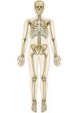
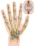
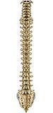
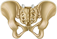
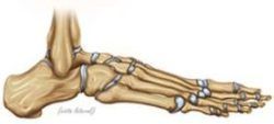
Skeletal System Anatomy. These illustrations are focused on the human skeletal system. Essentially, this structure supports the most important internal organs and soft tissues. We can also say that the skeleton is the set of bony parts that provide the human body with a firm, multifunctional structure—with the exception of the hyoid bone, which is separate from the rest of the skeleton. Therefore, all other bones are articulated with one another and supported by complementary connective structures such as ligaments, tendons, muscles, and cartilage.
You can also customize the image to suit your needs; the surcharge will depend on its complexity.
You can also request a custom illustration tailored to your needs; the surcharge will depend on its complexity.
The images are linked to their respective descriptions and, of course, to the buy button.


___________________________________________________________________________________________________________________________


__________________________________________________________________________________________________________________________


__________________________________________________________________________________________________________________________


__________________________________________________________________________________________________________________________



___________________________________________________________________________________________________________________________________________



___________________________________________________________________________________________________________________________________________




___________________________________________________________________________________________________________________________________________


_____________________________________________________________________________________________________________________________________________

___________________________________________________________________________________________________________________________________________
Bone structures
Illustrations Skeletal System. In these illustrations, we can see that the human bone structure is made up of 206 bones, 80 axial bones, among which are the bones of the head, face, ribs, and sternum. Also, 126 appendicular bones, among which are: arms, shoulders, wrists, hands, legs, and feet. The bone skeleton is a structure typical of vertebrates. For this reason, in Biology, the skeleton is any rigid or semi-rigid structure that provides support and also provides the basic morphology of the body; some facial cartilages (nasal, auricular, etc.) should also be considered part of the skeleton.
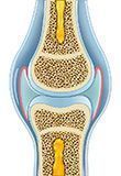
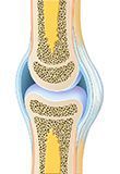
First of all, we have 2 different types of bones, compact and spongy. Compact bones form the outer and hardest layer of bones; this type of bone makes up 80% of bone mass. They also provide protection, support, and resist the efforts that occur during movements. Next, the spongy bones form the majority of the vertebral body; they consist of a network called trabeculae. Within each trabecula are cells that directly receive nutrients from the blood that circulates through our body.
